Skeleton Back Bones Diagram : Labeled Skeleton Back View Of Male Skeleton Anatomy Human Anatomy Male Skeleton / This diagram depicts back skeletal anatomy with parts and labels.
Skeleton Back Bones Diagram : Labeled Skeleton Back View Of Male Skeleton Anatomy Human Anatomy Male Skeleton / This diagram depicts back skeletal anatomy with parts and labels.. Human skeleton, the internal skeleton that serves as a framework for the body. Bones of the back anatomy tutorials. Bones of the pelvis and lower back. Skull bones protect the brain and form an entrance to the body. Every skeletal muscle has three main parts:
The lumbar spine connects to the thoracic spine above and the hips below. The bones of the appendicular skeleton (the limbs and girdles) append to the axial skeleton. The bones of the appendicular skeleton provide support and flexibility at the joints and anchor the muscles that move the limbs. These bones are connected at the back with specialized joints. Spinal anatomy and back pain.
Back Of The Human Skeleton Body Anatomy From Different Point Of View Bones Reference For Persons Posterior Medical Canstock from comps.canstockphoto.com Spinal vertebrae bone spine vertebra toracica spinal cord spine structure back diagram spine sections spinal cord vertebrae spinal structure health diagram. On the back of each vertebra are bony projections that form the vertebral arch. The vertebral column is a part of the axial skeleton, which comprises the skull, ribs and sternum other than the vertebral column. Human arm bones labeled, human hand bones labeled, human skeleton labeled back view, human skeleton labeled pdf, labeled bones in the human body, bone, human arm bones labeled, human. Bones of the pelvis and lower back. They support bones, in this case, the vertebrae. Anatomical diagrams of the spine and back. Pcs, handys, zubehör & mehr
Individual anatomical structures include 2:
A posterior view skeletal diagram provides a back view of the human skeleton. Skeleton back bones diagram / human skeleton anatomy vintage 1940s high res digital image / in this assignment, students color the various parts of the skeletal system and then answer some follow up teach your students the names of the bones in the human body with the help of this illustrated human skeleton diagram. The bones of the appendicular skeleton provide support and flexibility at the joints and anchor the muscles that move the limbs. The cranial bones compose the top and back of the skull and enclose the brain. Spinal anatomy and back pain. Bone tissue anatomy and physiology 12 photos of the bone tissue anatomy and physiology anatomy and physiology bone tissue test, anatomy and physiology chapter 6 skeletal system bone tissue, anatomy and physiology osseous tissue and bone structure quiz, bone anatomy and physiology ppt, bones anatomy and physiology test, bone, anatomy and. Anatomical diagrams of the spine and back. Skull bones protect the brain and form an entrance to the body. Because of this, they are referred to as cause and. The axial skeleton is made up of the skull, backbone, breastbone, and ribs. They support bones, in this case, the vertebrae. Human arm bones labeled, human hand bones labeled, human skeleton labeled back view, human skeleton labeled pdf, labeled bones in the human body, bone, human arm bones labeled, human. Bones of the pelvis and lower back.
The bones of the appendicular skeleton provide support and flexibility at the joints and anchor the muscles that move the limbs. This diagram depicts back skeletal anatomy with parts and labels. Spinal anatomy and back pain. The cranial bones compose the top and back of the skull and enclose the brain. A posterior view skeletal diagram provides a back view of the human skeleton.
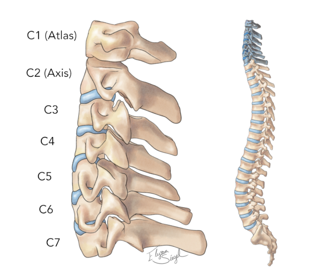
Spine Anatomy Goodman Campbell from www.goodmancampbell.com The vertebrae, which stack like spools of thread, support the back and protect the spinal cord. It is also known as the vertebral column. The spine or backbone consists of 26 small bones or vertebrae. Browse 2,180 human skeleton diagram stock photos and images available, or start a new search to explore more stock photos and images. The hollow spinal canal contains the spinal cord, fat, ligaments, and blood vessels. Browse 222 lower back skeleton stock photos and images available, or start a new search to explore more stock photos and images. The bones of the appendicular skeleton provide support and flexibility at the joints and anchor the muscles that move the limbs. The vertebral column is a part of the axial skeleton, which comprises the skull, ribs and sternum other than the vertebral column.
The bones of the pelvis and lower back work together to support the body's weight, anchor the abdominal and hip muscles, and protect the delicate vital organs of the vertebral and abdominopelvic cavities.
The vertebral column is a part of the axial skeleton, which comprises the skull, ribs and sternum other than the vertebral column. Skull bones protect the brain and form an entrance to the body. Bone tissue anatomy and physiology 12 photos of the bone tissue anatomy and physiology anatomy and physiology bone tissue test, anatomy and physiology chapter 6 skeletal system bone tissue, anatomy and physiology osseous tissue and bone structure quiz, bone anatomy and physiology ppt, bones anatomy and physiology test, bone, anatomy and. Anatomical diagrams of the spine and back. The spine or backbone consists of 26 small bones or vertebrae. Skeletal diagrams can also be used to show bone development or growth which begins en utero. The vertebral column of the lower back includes the five lumbar vertebrae, the sacrum, and the coccyx. Learn vocabulary, terms and more with flashcards, games and other study tools. Spinal vertebrae bone spine vertebra toracica spinal cord spine structure back diagram spine sections spinal cord vertebrae spinal structure health diagram. The arch is made of two supporting pedicles and two laminae (fig. The axial skeleton is made up of the skull, backbone, breastbone, and ribs. The bones of the appendicular skeleton (the limbs and girdles) append to the axial skeleton. This article looks at the anatomy of the back, including bones, muscles, and nerves.
Skeletal diagrams can also be used to show bone development or growth which begins en utero. Spinal anatomy is a remarkable combination of strong bones, flexible ligaments and tendons, large muscles and highly sensitive nerves. Everything else that hangs from this, like the arms, legs, shoulders, and hips, is called the appendicular skeleton. Human back bones diagram poster 28 inch x 24 inch 16 inch x 13 inch. The spine anatomy is a complex structure.
Human Being Anatomy Skeleton Posterior View Image Visual Dictionary from www.ikonet.com This human anatomy module is composed of diagrams, illustrations and 3d views of the back, cervical, thoracic and lumbar spinal areas as well as the various vertebrae. The lumbar spine connects to the thoracic spine above and the hips below. Human skeleton, the internal skeleton that serves as a framework for the body. This article looks at the anatomy of the back, including bones, muscles, and nerves. The hollow spinal canal contains the spinal cord, fat, ligaments, and blood vessels. It is particularly interesting for physiotherapists. And coccygeal the tail bone. They support bones, in this case, the vertebrae.
By tightening and relaxing, the skeletal muscles create movement.
And coccygeal the tail bone. The bones of the skeletal system act as attachment points for the skeletal muscles of the body. Bones of the pelvis and lower back. The lumbar spine connects to the thoracic spine above and the hips below. Spinal anatomy and back pain. The vertebral column of the lower back includes the five lumbar vertebrae, the sacrum, and the coccyx. The red lines point individual bones and the names are writen in singular, the blue lines conect to group of bones and are in plural form. Human skeleton, the internal skeleton that serves as a framework for the body. Your lower back contains 5 vertebral bones stacked above each other with intervertebral discs in between. It also covers some common conditions and injuries that can affect the back. Diagram showing the bones of the human arms and shoulders, circa 1900. Découvrez tout de suite de nouvelles offres de nos dernières gammes chez boohooman. *the origin, insertion, and belly.* a muscle's origin is where a tendon attaches it to the *less* movable bone.
Every skeletal muscle has three main parts: back bones diagram. Spinal vertebrae bone spine vertebra toracica spinal cord spine structure back diagram spine sections spinal cord vertebrae spinal structure health diagram.


0 Komentar